
 Do you feel frightened on seeing threatening images? As per a study published in the journal Brain and Cognition, researchers from the Center for BrainHealth at the
 Scientists analysed previous animal and human research to find an electrophysiological marker for threat in the brain.
According to
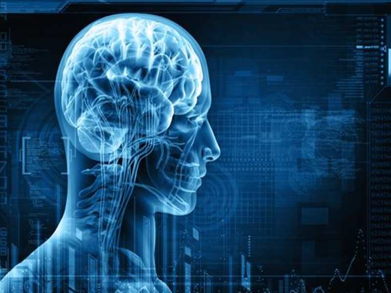
Utilising electroencephalography (EEG), the research team led by Hart identified theta and beta wave activity that signifies the brain's reaction to visually threatening images.
"We have known for a long time that the brain prioritises threatening information over other cognitive processes," said
"These findings show us how this happens. Theta wave activity starts in the back of the brain, in it's fear center — the amygdala — and then interacts with brain's memory center — the hippocampus — before travelling to the frontal lobe where thought processing areas are engaged. At the same time, beta wave activity indicates that the motor cortex is revving up in case the feet need to move to avoid the perceived threat," said DeLaRosa.
In order to conduct the study, 26 adults (19 female, 7 male) falling in the age bracket of 19-30 were shown 224 randomised images that were either unidentifiably scrambled or real pictures. Real pictures were separated into two categories: threatening (weapons, combat, nature or animals) and non-threatening (pleasant situations, food, nature or animals).
While wearing an EEG cap, participants were asked to press a button with their right index finger for real items and another button with their right middle finger for nonreal/scrambled items. Shorter response times were recorded for scrambled images than the real images. There was no difference in reaction time for threatening versus non-threatening images.
The results of the study showed that threatening images evoked an early increase in theta activity in the occipital lobe, the area in the brain where visual information is processed. This was followed by a later increase in theta power in the frontal lobe, the area of brain where higher mental functions such as thinking, decision-making and planning occur.
So know you the reason that why you feel frightened on seeing threatening images.
(Image: Thinkstock)