These 20 winning microscope images reveal a beautiful, hidden universe in incredible detail
20) Fungus growing on cow dung

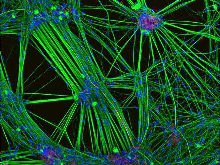
19) Brain cells grown from human embryonic stem cells
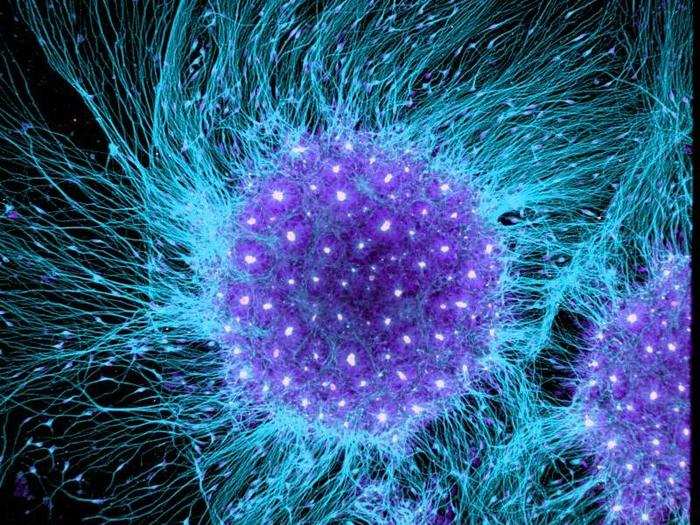
Six Rockefeller University researchers snapped this photo (magnified 10X) of embryonic stem cells coaxed to grow into brain cells.
"We use human stem cells to model and understand human brain development and diseases such as [Huntington's] and Alzheimer's disease," the group wrote in its photo entry.
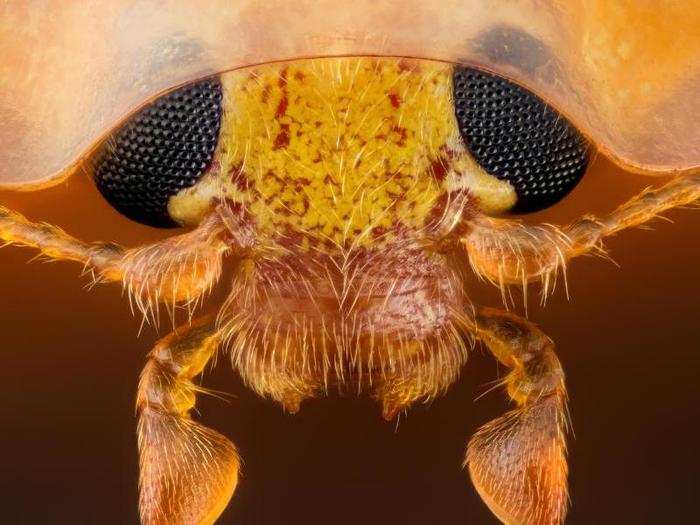
18) Hind leg of a broad-shouldered leaf beetle
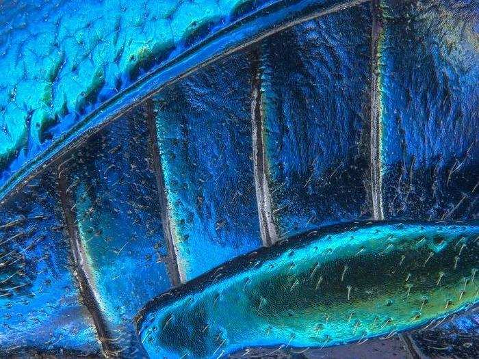
This image (magnified 40X) isn't just for beauty, says its maker — Pia Scanlon, a bio-security specialist in the Department of Agriculture and Food in Western Australia.
"My work requires me to understand and capture taxonomic detail while creating an appealing image," she wrote in her photo entry. "Often it is important, from a taxonomic point of view, to microphotograph living [specimens] prior to curation to capture the full colour spectrum of a species. This gives an accurate reproduction of how an insect look’s in its natural environment. Preserved specimens can lose their colors over time, for example the eye colour of dragonflies diminishes almost as soon as they die."
17) Slime mold
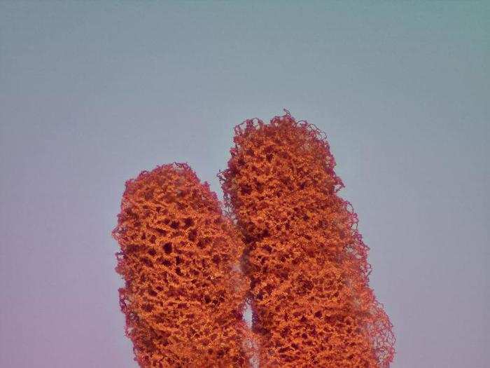
Jose Almodovar, a biologist at the University of Puerto Rico, found this mold growing in a field.
The two fruiting bodies look like "kissing people at this scale" of 5X magnification, he wrote in his photo entry.
16) Zooplankton carefully arranged by hand in a Victorian style
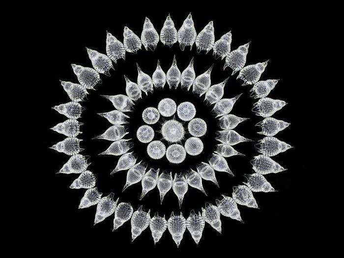
"Plankton in general have often artistic shapes that lead us to reflect upon spiritual matters and wonders of Nature," Italian photographer Stefano Barone wrote in his entry for this photo (magnified 100X).
He said Radiolarians — the type of plankton, or ocean organisms, that he arranged in the image — "emanate hypnotic beauty, magic, mathematics, mystery, fun and solemnity."
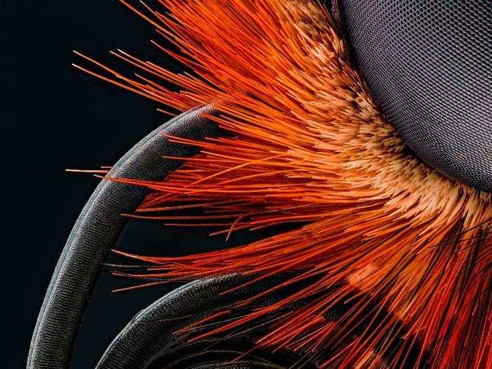
15) Head of an orange ladybird beetle
Electronics engineer Geir Drange took this shot, magnified 10X.
"I frequently encounter these beetles in my moth trap, as they are drawn towards light," Drange wrote in his photo entry. "It shows interesting details of this little beetle, like the beetle's shield is translucent."
14) Retinal nerve cells from a mouse eye
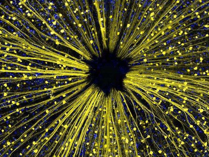
In his entry for this photo (magnified 40X), Dr. Keunyoung Kim, a researcher at the University of California, San Diego, said this mouse retina "appeared to embody a galaxy of infinite secrets and endless potential for discovery. The optic disc area of the retina also fascinated me, with its retinal ganglion cells and axon's array resembling the sun's rays."
13) Poison fangs of a centipede
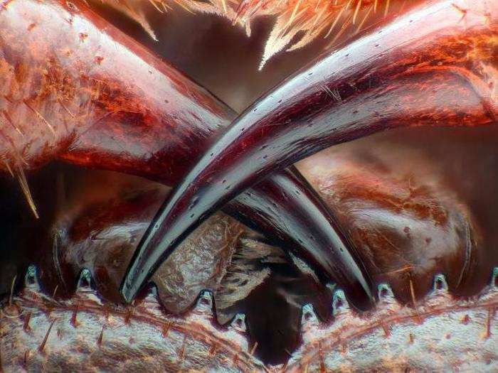
"The image shows the fangs that deliver a poison to kill the prey of the centipede," photographer Walter Piorkowski of South Beloit, Illinois, wrote in his entry. "In addition the 10 coxosternal teeth are also visible. Fine feather like hairs or setae in front of the mouth and above the fangs around the first maxilae."
"Personally, the centipede has always been a [subject] of unease due to their look and remarkable speed," he said of the image (magnified 16X).
12) Human HeLa cell undergoing cell division
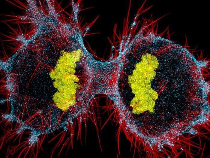
Dr. Dylan Burnette, a researcher at Vanderbilt University School of Medicine, submitted this image, writing in his entry:
"This image is of a cancer cell in the final stage of cell division where one 'mother' cells become [sic] two 'daughter' cells. This process drives the ability of tumors to grow as well as the ability of body to regenerate organs and tissue. We are interested in how the two cells get physically separated by the so called 'cleavage furrow'. The cleavage furrow is the pinch point in between the two cells, and constricts to until two cells are separated. Our research is focused on using superresolution [microscopy] to investigate how the molecular machines driving the constriction of the cleavage furrow are assembled. One such machine, myosin II, can be seen in blue."
11) Scales from the underside of a butterfly wing
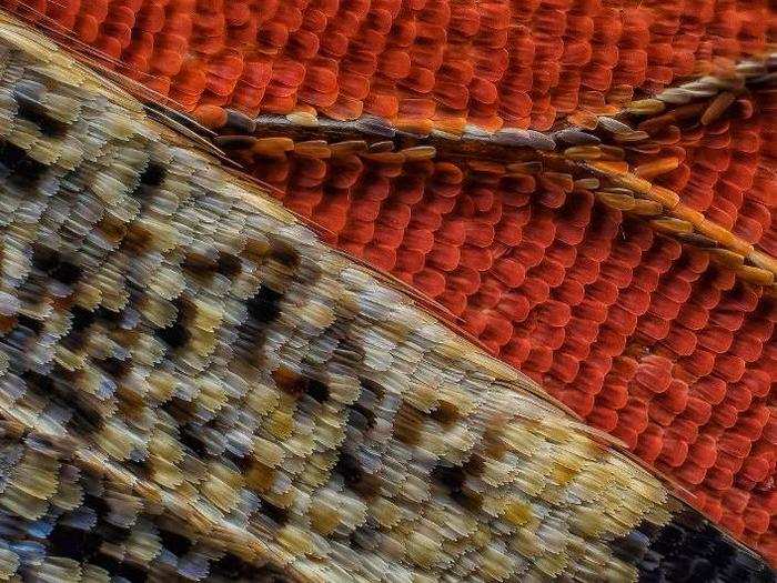
"It is interesting to sea that these scales are very [similar] to scales of a fish. The big difference is that the scales of a butterfly are extremely vulnerable, and will break off when touched with your fingers," photographer Francis Sneyers of Belgium wrote about this photo (magnified 10X) in his entry.
10) A single-celled protist showing its ingested food, cilia, mouth, and trichocysts
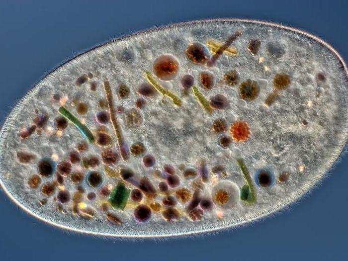
Rogelio Moreno Gill of Panama says this 200X-magnified image of a single-celled Frontonia protist looks more "like a pizza" than a microbe.
"This image shows many details of the Frontonia: the mouth (at the lower right side), the cilia (around the Frontonia), the ingested foods (all the items with colors that we see inside the Frontonia) and the Trichocysts (around the internal part of the Frontonia)," Gill wrote in his photo entry.
9) Espresso coffee crystals
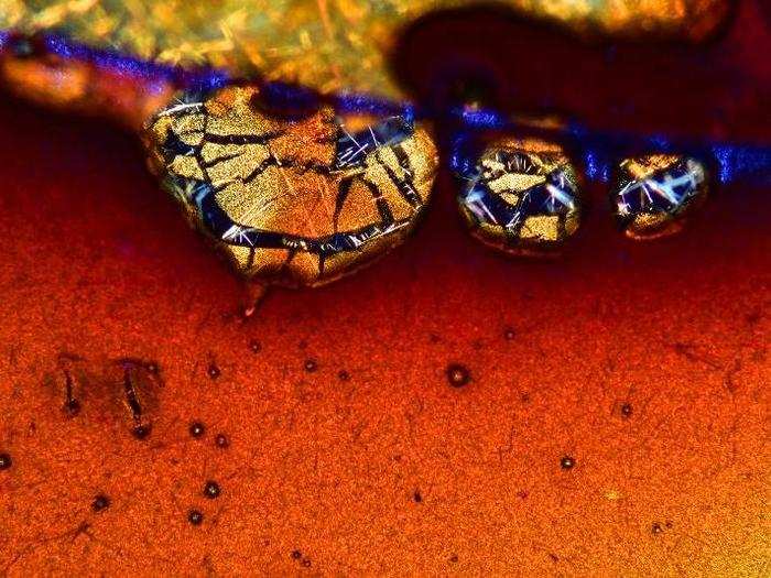
The photomicrograph was taken by artist and environmental scientist Vin Kitayama, who runs the Vinsanchi Art Museum in Japan, and his wife Sanae Kitayama.
"During my research, I discovered the mystery and beauty of natural design that is hidden in one small drop of coffee," Vin wrote in his photo entry. "The natural gold color in this photograph reminds me of the beautiful gold that you sometimes can see in the finest traditional Japanese lacquer work, such as created by the famous artist, Korin, about three hundred years ago."
The couple said the technique they used to take this photo was "developed over a long period" and took "a most difficult process" to get the espresso to crystallize. (Business Insider contacted Vin for more details, but he did not immediately respond.)
You can see filaments of crystallized chemicals — perhaps caffeine, which is white as a pure crystal — propping up cracked, golden blobs of cream, or coffee foam.
8) Wildflower stamens
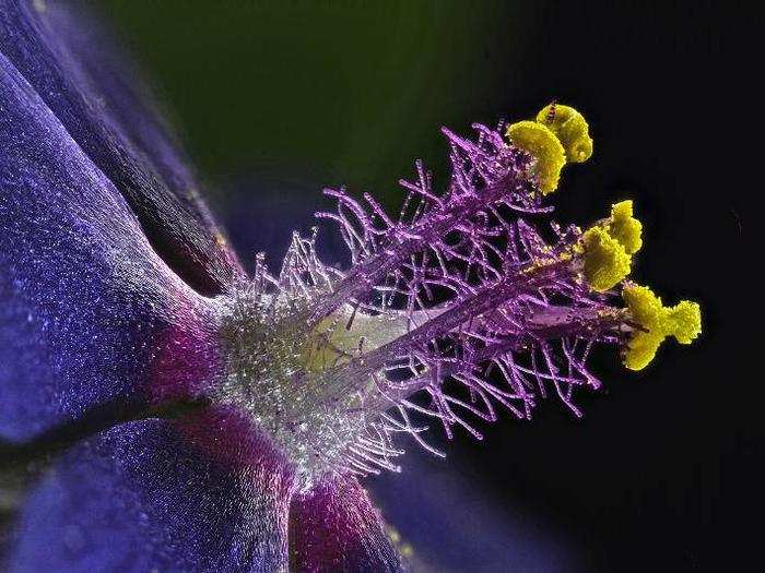
Israel-based artist Monoson Yahud, a microscope photographer of 20 years, merged 100 different photos to create this image (magnified 40X).
"Only through a microscope," Yahud wrote in his photo entry, can you see a flower's true beauty.
7) Leaves of a lesser club moss
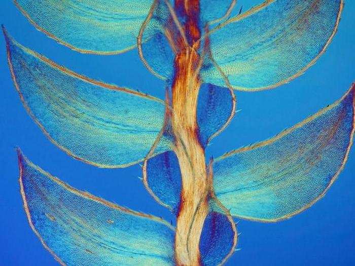
Zoologist, photographer, and journalist David Maitland took this image, which is magnified 40X. "I have a passion for invertebrates and plants — especially the aesthetic between form and function the sheer beauty of nature," he wrote in his photo entry. "The majority of life on earth is tiny, and often invisible."
6) Melted vitamin C crystals

A biologist by education, Marek Mis took this image (magnified 50X) after melting down ascorbic acid — known better as vitamin C — to find a rainbow-colored assortment of air bubbles.
5) Front foot of a male diving beetle
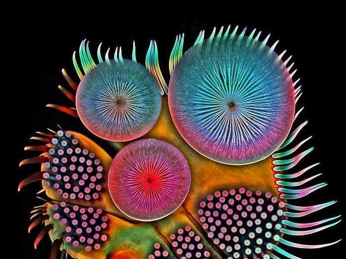
Although Igor Siwanowicz, a research scientist at Howard Hughes Medical Institute, normally studies the brains of dragonflies as they capture prey, he told Nikon that he's captivated by other invertebrate animals, too.
To that end this image shows the front foot, or tarsus, of male diving beetle and its clusters of suction cups — devices the insect uses to stick to a female beetle for mating.
4) Butterfly proboscis, or tongue
Thailand-based micro-photographer Jochen Schroeder took this photo of a butterfly's mouth, which is actually a bunch of separate images merged together to reveal more detail.
3) Brain cells grown from human skin cells
This image from Rebecca Nutbrown, a neuroscience PhD student at University of Oxford, shows two different types of brain cells and their axons, or nerve fibers: neurons (stained green) grown from human skin cells, and Schwann cells (stained red) grown from a rodent. The cell bodies are stained blue.
"I am fascinated by how the microscope and fluorescence can reveal such complex beauty, completely missed by the naked eye," Nutbrown said in a Nikon press release.
2) Polished slab of Teepee Canyon agate
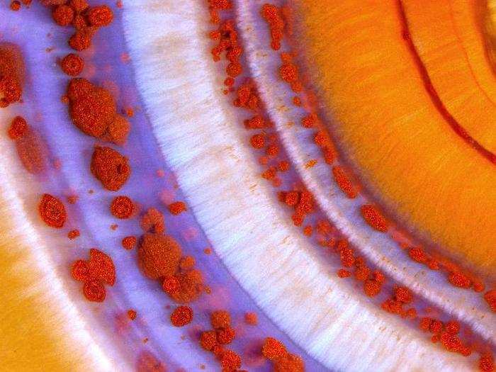
Seen at 90X magnification, this Teepee Canyone Agate is a 273-million-year-old slice of ocean sediments recovered from the Black Hills of western South Dakota.
Specimens like this "sometimes contain fossils or are replacements of fossil structures such as coral heads or sponges," according to Nikon.
1) Four-day-old zebrafish embryo
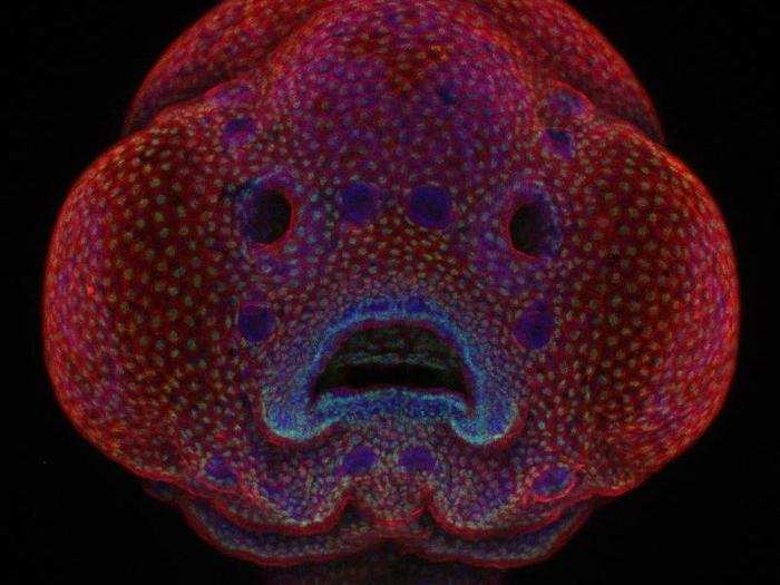
Oscar Ruiz, a researcher at the MD Anderson Cancer Center in Houston, Texas, took this photo of a four-day-old zebrafish embryo — and it won the Nikon Small World contest's grand prize.
Ruiz says he takes advantage of the zebrafish's translucent face to study "genetic mutations that lead to cleft palate and cleft lip in humans," according to a Nikon press release.
His ultimate goal? To build up an atlas of facial development that might "provide insight and lead to solutions for preventing and correcting facial deformities in people in the near future."
Popular Right Now
Advertisement