The best microscope photos of 2022 reveal a hidden world of dino-bone crystals, human tongue bacteria, and slime mold
Morgan McFall-Johnsen

The winning photo from the 2022 Nikon Small World competition: the hand of a gecko embryo.Grigorii Timin & Dr. Michel Milinkovitch/Nikon Small World
- Nikon's Small World competition recognizes the best microscope photographs of the year.
- Microscopy is an art and a science, revealing the alien beauty of the hidden world all around us.
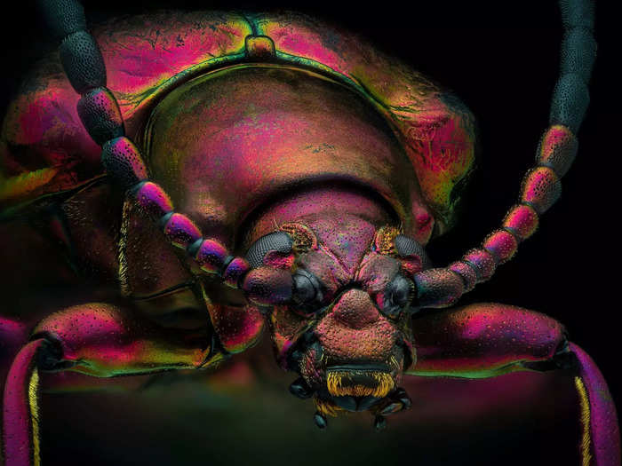
The Nikon Small World competition recognizes the art and science of microscope photography, or "microscopy."
Yousef Al Habshi/Nikon Small World
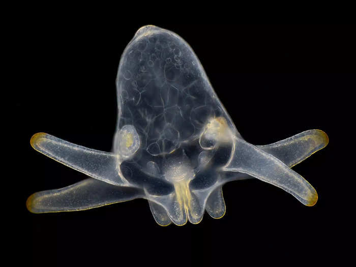
Scientists, artists, and enthusiasts from all over the world submit their painstakingly crafted photos of cells, nerves, micro crystals, mold, and tiny creatures like this anemone larva.
Wim van Egmond/Nikon Small World
This year, Grigorii Timin won first place by stitching together hundreds of images to reveal the nerves, bones, tendons, ligaments, and blood cells in the 3-millimeter-wide hand of a gecko embryo.
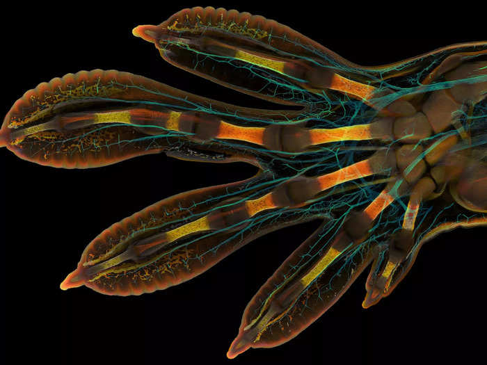
The winning photo from the 2022 Nikon Small World competition: the hand of a gecko embryo. Grigorii Timin & Dr. Michel Milinkovitch/Nikon Small World
That's a Madagascar giant day gecko. For reference, this is what they look like at full size.
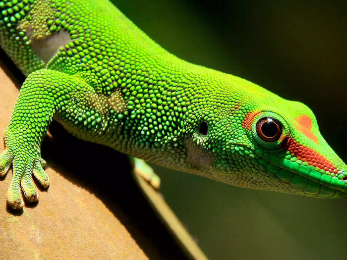
A Madagascar day gecko sits on a perch in the Masoal rainforest hall at the zoo in Zurich, on March 19, 2013. Arnd Wiegmann/Reuters
Second place went to this close-up photo of breast tissue, showing cells wrapped around the alveoli that produce milk.
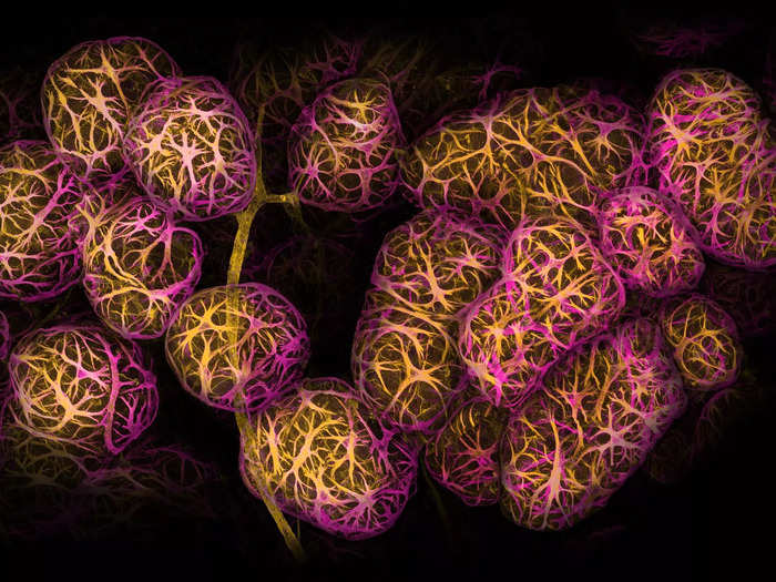
Dr. Caleb Dawson/Nikon Small World
The otherworldly blood vessel networks in a mouse's intestine took third place.
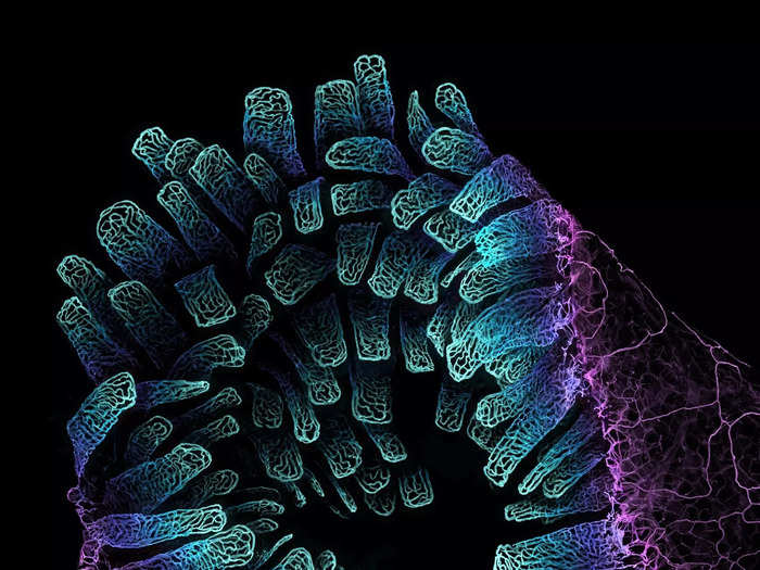
Satu Paavonsalo & Dr. Sinem Karaman/Nikon Small World
A portrait of a daddy longlegs spider snagged fourth place.
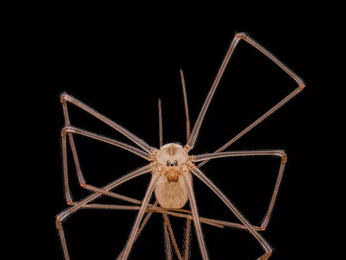
Dr. Andrew Posselt/Nikon Small World
Here are the rest of the 20 winners:
5. Slime mold is a common microscopy subject, since it grows in eerie formations of colorful bulbs.
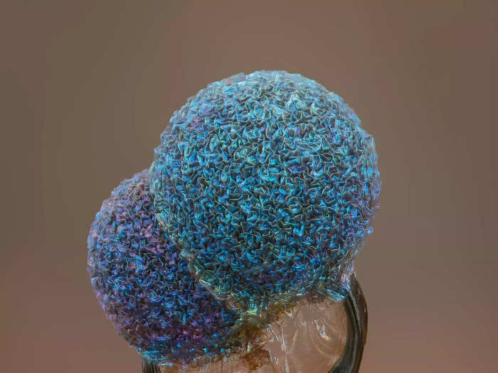
Alison Pollack/Nikon Small World
6. Unburned particles of carbon, released as the hydrocarbon chain of candle wax breaks down.
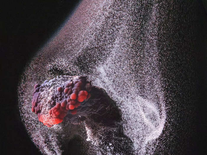
Ole Bielfeldt/Nikon Small World
7. Neurons from human neural stem cells.
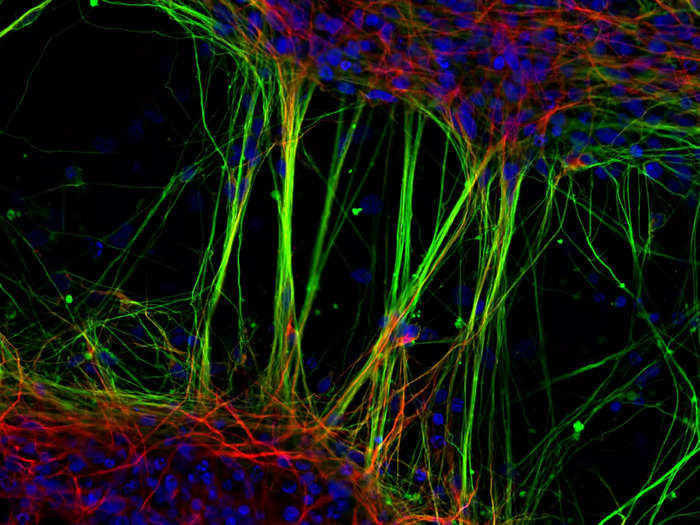
Dr. Jianqun Gao & Prof. Glenda Halliday/Nikon Small World
8. The growing tip of a frond of red algae.
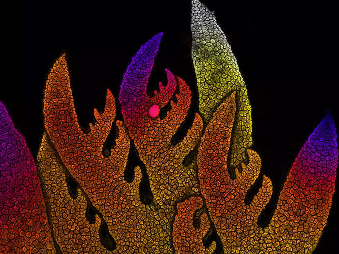
Dr. Nathanaël Prunet/Nikon Small World
9. A mixture of liquid crystal, which resembles a human portrait.
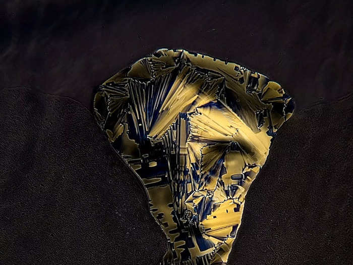
Dr. Marek Sutkowski/Nikon Small World
10. A fly under the chin of a tiger beetle.
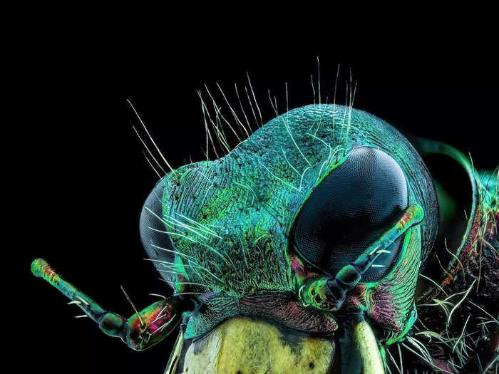
Murat Öztürk/Nikon Small World
11. A stack of moth eggs.
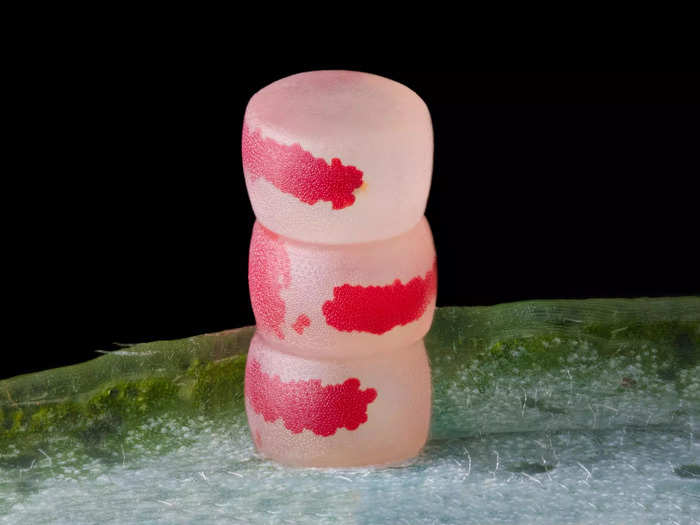
Ye Fei Zhang/Nikon Small World
12. A single polyp of coral, about 1 millimeter wide, giving off fluorescence.
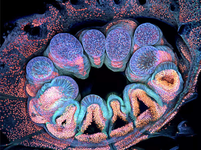
Brett M. Lewis/Nikon Small World
12. Agate — a colorful, crystallized quartz — that formed inside a dinosaur bone when it was infiltrated with silica-rich water.
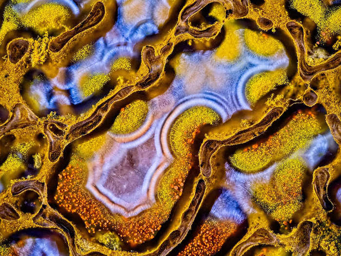
Randy Fullbright/Nikon Small World
14. Mouse myoblasts — precursors of the cells that build muscle tissue — with their nuclei in yellow, lysosomes in cyan or green, and filaments in magenta.
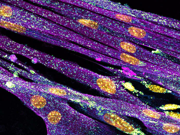
Nadia Efimova/Nikon Small World
15. Cross sections of cells from the lining of a human colon, which look like a sea of flowers.
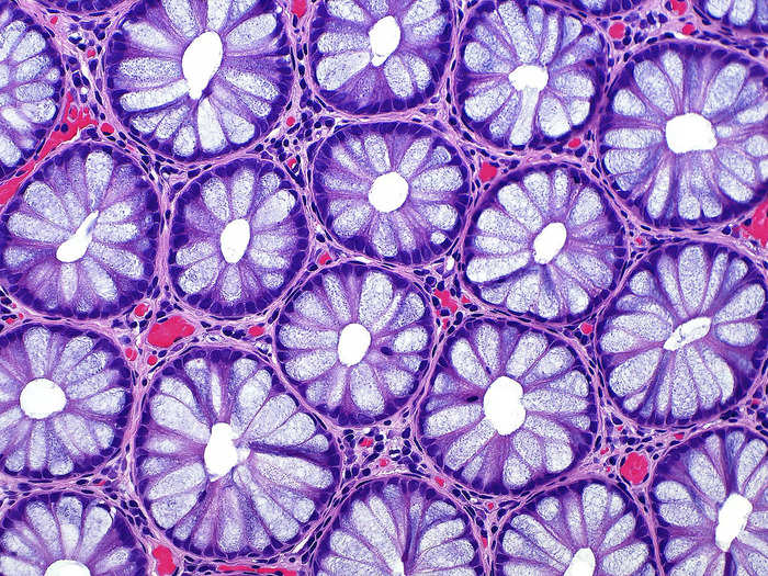
Dr. Ziad El-Zaatari/Nikon Small World
16. A section cut out of the top of a shoot of white asparagus.
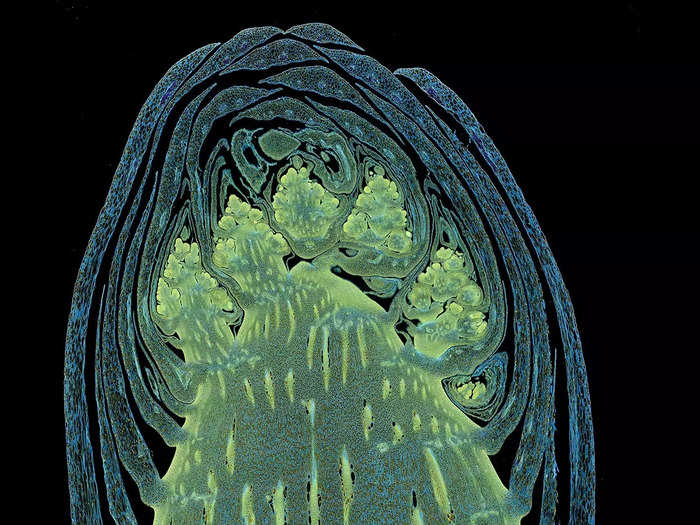
Dr. Olivier Leroux/Nikon Small World
17. A zebrafish larva's tail fin, with peripheral nerves marked green and the structure around cells marked violet.
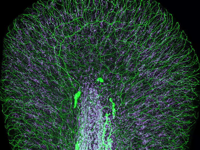
Dr. Daniel Wehner & Julia Kolb/Nikon Small World
18. A network of white blood cells in a zebrafish intestine.
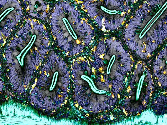
Dr. Julien Resseguier/Nikon Small World
19. A "biofilm" of bacteria on a human tongue cell.
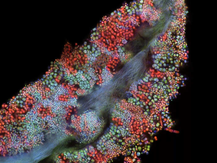
Dr. Tagide deCarvalho/Nikon Small World
20. Human heart cells, captured at the Nationwide Children's Hospital Center for Cardiovascular Research.
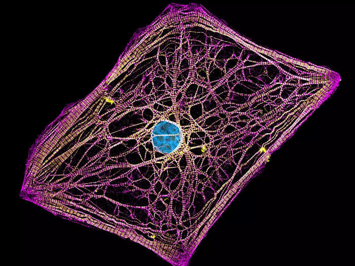
Hui Lin & Dr. Kim McBride/Nikon Small World
Popular Right Now
Popular Keywords
Advertisement