Award-Winning Images Of Some Of The Smallest Things On Earth
First place winner, Mr. Wim van Egmond, of the Micropolitan Museum in The Netherlands took this image of a marine diatom called Chaetoceros debilis, which lives as a colony. It is enlarged 250 times.

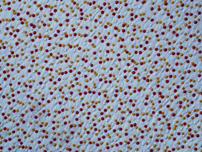
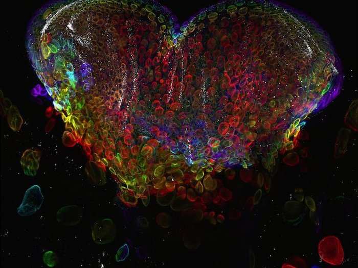
Dr. Joseph Corbo, of Washington University School of Medicine, took this image of the retina of the painted turtle (Chrysemys picta). It is enlarged 400 times.
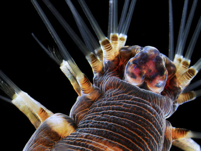
Dr. Alvaro Esteves Migotto of the Universidade de Sa~o Paulo in Brazil took this image of a marine worm, enlarged 20 times.
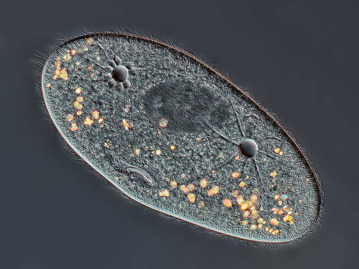
Mr. Rogelio Moreno Gill of Panama City, Panamá took this image of a species of Paramecium showing the nucleus, mouth, and water expulsion vacuoles, enlarged 40 times.
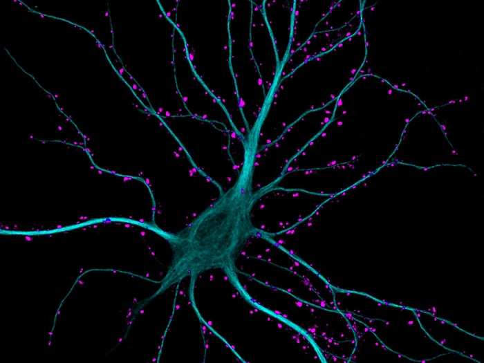
Dr. Kieran Boyle of the University of Glasgow, in Scotland took this image of a brain cell in the hippocampus, receiving excitatory contacts. It is enlarged 63 times.
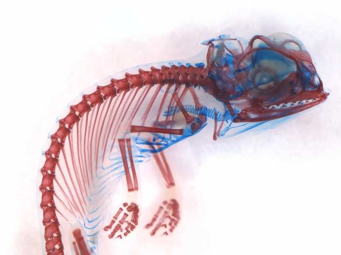
Miss Dorit Hockman, of the University of Cambridge in the U.K., took this image of a veiled chameleon (Chamaeleo calyptratus) embryo that has been stained to highlight the cartilage (blue) and bone (red). The rest of the tissues have been made clear.
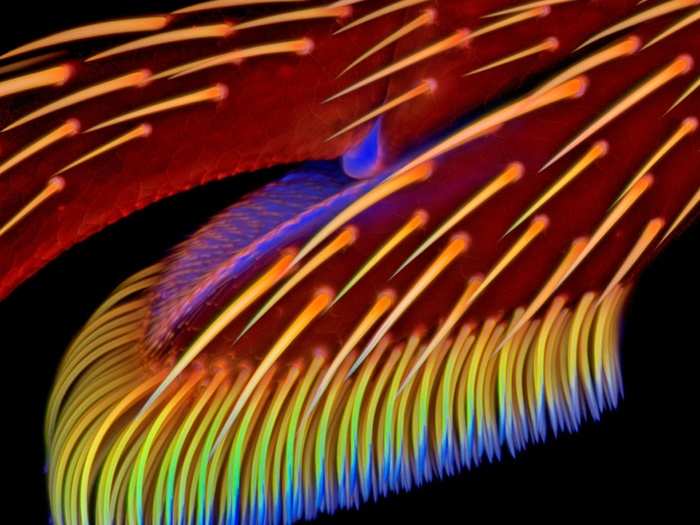
Dr. Jan Michels of the Institute of Zoology at Christian-Albrechts-Universität zu Kiel in Germany took this image of the adhesive pad on a foreleg of a ladybird beetle (Coccinella septempunctata) also known as a ladybug. It's enlarged 20 times.
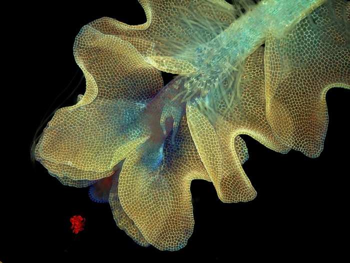
Ms. Magdalena Turzan´ska, of the University of Wroclaw in Poland, snapped this image of a liverwort (not liverworst) plant of the species Barbilophozia and the cyanobacteria on it that fluoresce under UV light. It has been enlarged 50 times.
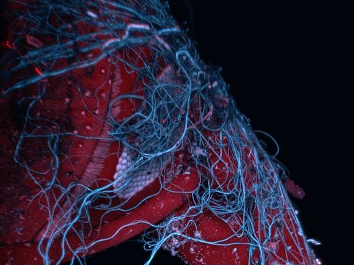
Mr. Mark A. Sanders of the University of Minnesota took this image of an insect wrapped in spider web, which is magnified 85 times.
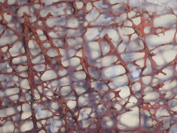
Mr. Ted Kinsman, of the Rochester Institute of Technology, took this image of a thin section of a dinosaur bone preserved in clear agate, enlarged 10 times.
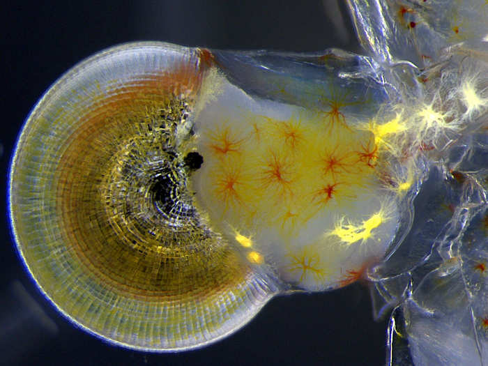
Miss Vitoria Tobias Santos, of the Universidade Federal do Rio de Janeiro in Brazil, took this image of a ghost shrimp (Macrobrachium) eye, magnified 140 times.
Dr. Pedro Barrios-Perez, of the National Research Council of Canada, took this image of silicon dioxide on polydimethylglutarimide-based resistor, magnified 200 times.
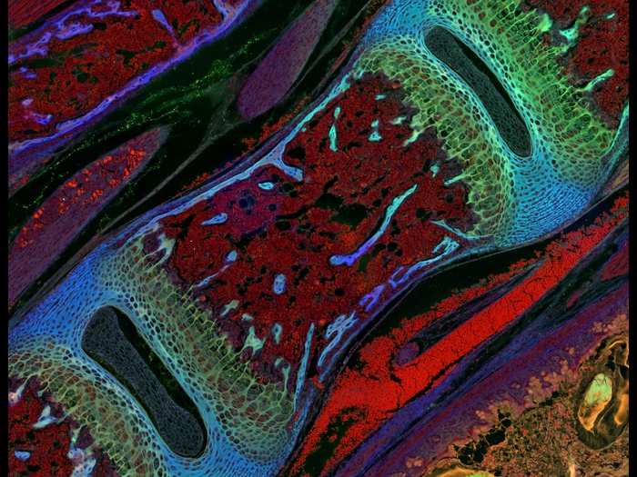
Dr. Michael Paul Nelson and Samantha Smith, of the University of Alabama at Birmingham, took this image of a section of the vertebra of a mouse, magnified 200 times.
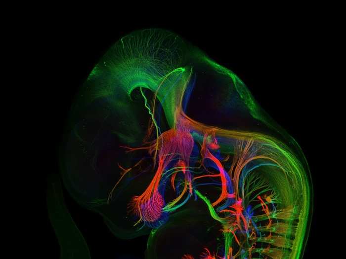
Mr. Zhong Hua, of Johns Hopkins University School of Medicine, took this image of the peripheral nerves (those outside of the brain) in an 11.5-day-old mouse embryo, magnified five times.
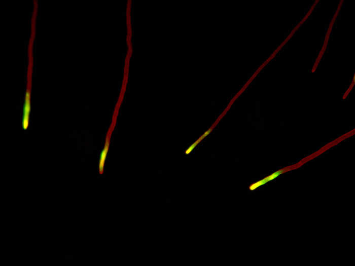
Dr. Christian Q. Scheckhuber, of Goethe University in Germany. took this image of the "filamentous tip cells" in the fungus Podospora anserina. The image was magnified 630 times.
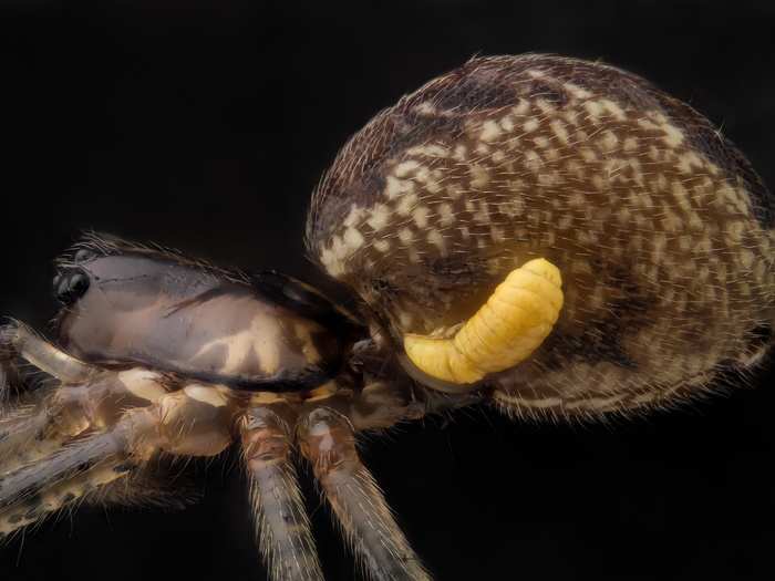
Mr. Geir Drange, of Asker, Norway, took this image of a sheet weaver spider (Pityohyphantes phrygianus) with a parasitic wasp larva on its abdomen, magnified five times.
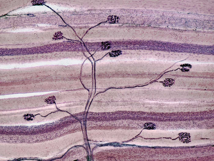
Dr. Alexandre William Moreau, of the University College London in the U.K., took this image of a special type of brain cell called pyramidal neurons, in the visual cortex of a mouse brain, enlarged 40 times.

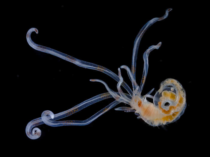
Mr. Christian Sardet, of the Centre National de la Recherche Scientifique in France, took this image of Annelid larva, magnified 100 times.
Dr. David Ward, of Oakdale, California, took this image of a thin section of nerve and muscle tissue. It is enlarged 40 times.
Dr. James Burchfield, of The Garvan Institute in Australia, took this image of a fat cell that shows the explosive dynamics of how the cell transports sugar.
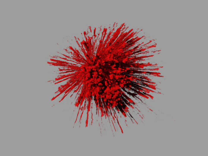
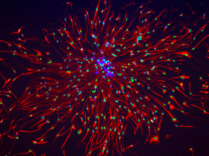
Honorable mention: Dr. Mariela Loschi, of Buenos Aires, Argentina, took this image of the microtubules and nucleus in a cultured cell, magnified 100 times.
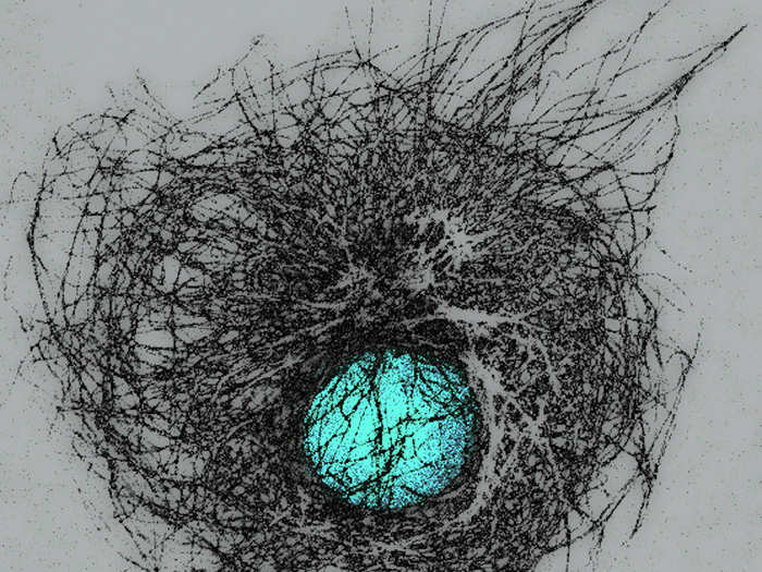
Honorable Mention: Dr. Michael J. Boyle, of the Smithsonian Institution, took this image of a peanut worm larvae magnified 40 times.
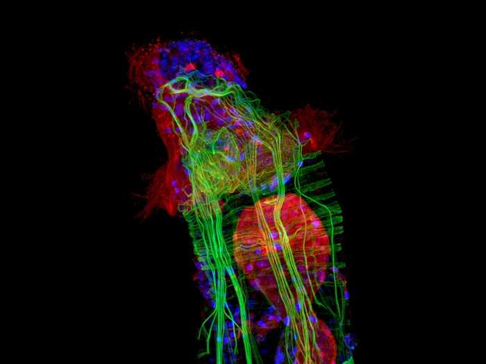
Image Of Distinction: Mr. Raul M. Gonzalez, of the Raul Gonzalez Studio in Mexico, took this image of a Vitamin C crystal, magnified 100 times.
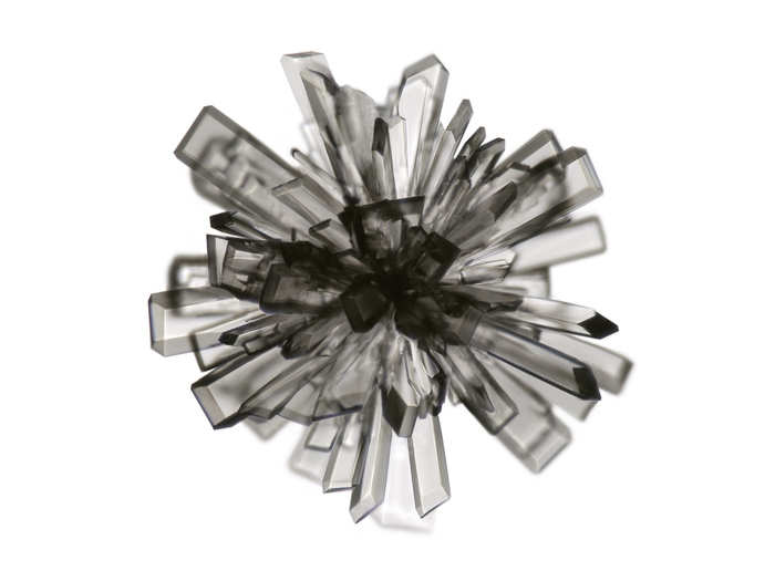
IOD: Dr. Biplab Dasgupta, Jane Anderson, Shabnam Pooya, Mariko DeWire, and Lili Miles, of Cincinnati Children’s Hospital and Medical Center, took this image of brain-cancer stem cells from human brain-cancer tissue, magnified 200 times.
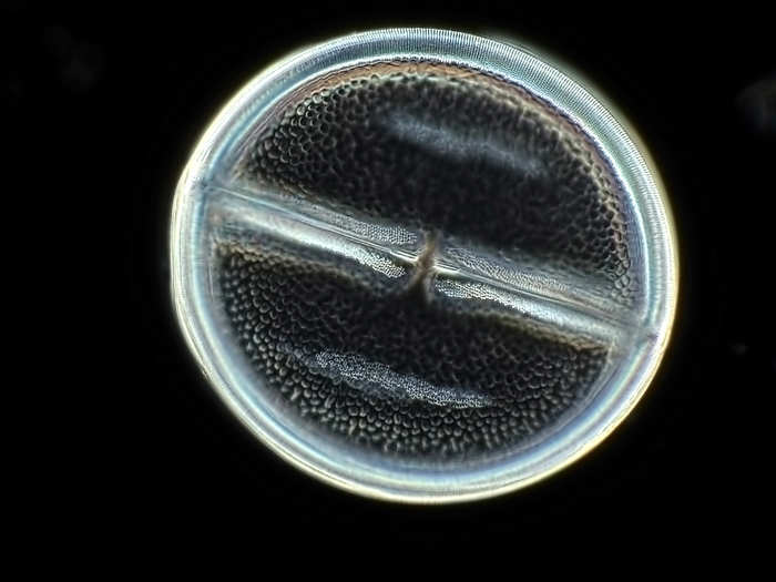
IOD: Mr. Arturo Agostino, of Reggio Calabria, Italy, took this picture of a tiny diatom called Navicula variolata, magnified 400 times.
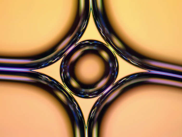
IOD: Mr. Haris Antonopoulos, of Athens, Greece, took this trippy image of a soap bubble magnified 10 times.
IOD: Dr. Guichuan Hou, of the Dewel Microscopy Facility in North Carolina, took this image of a cross section of lily of the valley (Convallaria) magnified 40 times.
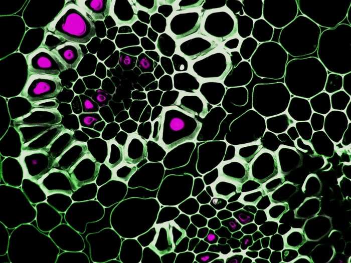
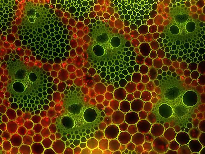
IOD: Mr. Eckhard Voelcker, of the Berlin Microscopic Society in Germany, took this image of the cross section of bamboo stems — which look decidedly spooked. They are magnified 200 times.
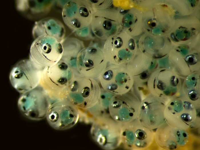
IOD: Dr. Jaime Gomez Gutierrez, of the Centro Interdisciplinario de Ciencias Marinas in Mexico, took this image of happy-looking fish eggs in a cluster. It's magnified 6.6 times.
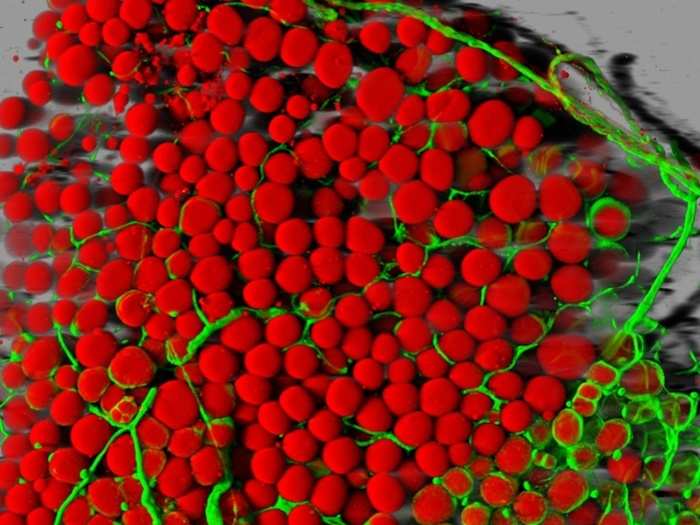
IOD: Dr. Daniela Malide, of the National Institutes of Health, took this picture of mouse fatty tissue. The red blobs are fat cells, and the green are blood vessels, magnified 25 times.
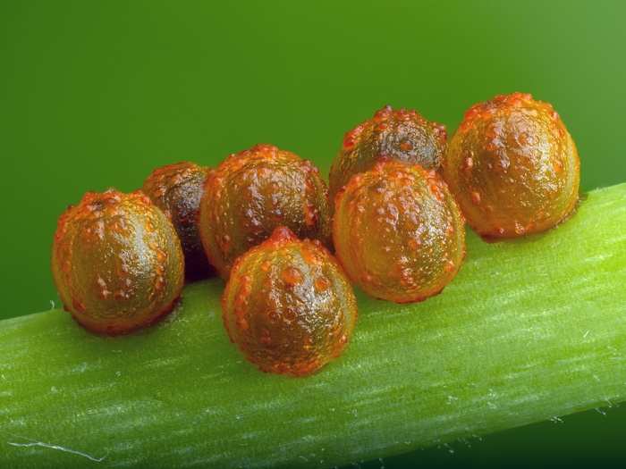
IOD: Mr. David Millard, of Austin, Texas, got this image of Pipevine Swallowtail butterfly (Battus philenor) eggs on stem of host plant, Aristolochia fimbriata, magnified five times.
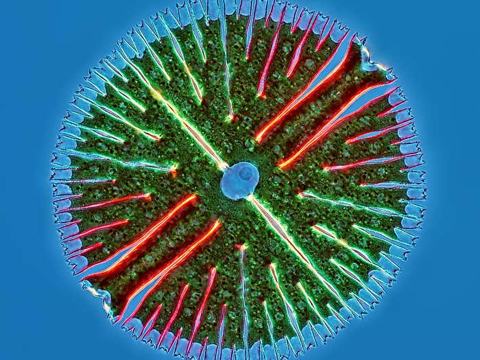
IOD: Mr. Rogelio Moreno Gill, of Panama City, Panama, took this picture of the algae Micrasterias radiosa, magnified 40 times.
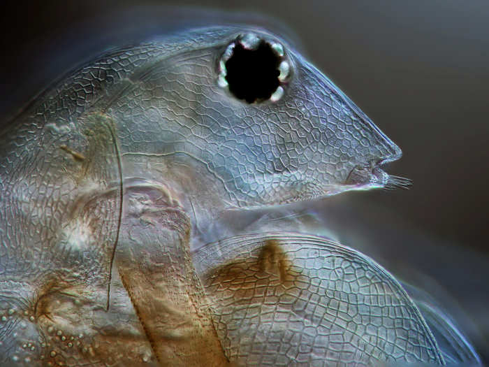
IOD: Mr. Jerzy Rojkowski, of Jerezy Rojkowski Photography in Poland, took this image of a confused-looking freshwater flea (Daphnia magna) magnified 200 times
See the images of microscopic life that won last year.
Popular Right Now
Popular Keywords
Advertisement