"Getting an ultrasound done used to be a destination," Martin said. There was a lot of waiting around for results, for technology, for the technicians and doctors to come proctor and administer these tests.
"It's time for the physical examination to change," Martin said.
Increasingly, medical schools and graduate medical education programs are incorporating ultrasound interpretation and proctoring into their curriculum. More healthcare providers are learning how to administer an ultrasound. These include nurses, nurse practitioners, EMTs, and other healthcare providers. Making a critical diagnosis out in the field or at remote locations ahead of entering the hospital can help providers on the ground anticipate what resources are needed for the patient ahead of time. The device has 13+ settings that can look at musculoskeletal, abdominal, aorta, bladder, cardiac, and pediatric abdomen. These indications are all FDA approved.
This device is not meant to completely phase out existing ultrasound machines, at least not as of now.
Martin said that they're to give healthcare providers a quick look at the significant issues such as general heart function or presence of a blood clot, so it can screen out people who don't need more complex tests.
"This is incredibly good for yes-no answers. Do you have an aneurysm or not?" Martin said. It's perfect as a bedside tool and as a portable device in developing countries with limited resources he added.
In fact, last year, Martin used the device to diagnose his own cancer.
As far as image resolutions go, the team said that in side-by-side comparisons with $60,000-$80,000 machines in the market right now, the difference is indiscernible.
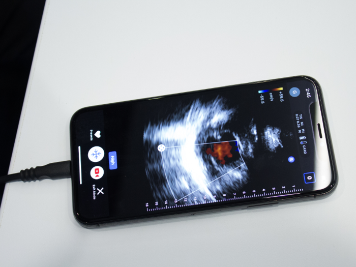
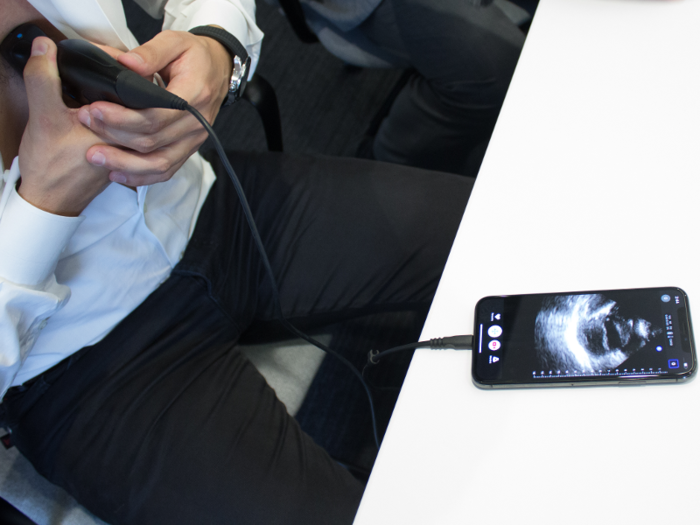
Darius Shahida, chief of staff at Butterfly, demonstrates the function of the cardiac setting of the ultrasound by imaging his own heart in real-time.
"You can see the valves and the contractility of the heart, so if someone comes in to the emergency room and I'm worried if their heart is functioning well or not," Martin said. "I can very quickly, with a very simple scan, within seconds, answer that question."
In conditions like pericardial effusion, where fluid gathers around the heart, a quick diagnosis can mean the difference between life or death. And sometimes there's not enough time to send for a technician, lug the heavy machinery to where the patient is. These are instances where a quick diagnosis and discharge for surgery can really make a medical difference.
The app also has advanced functionality like viewing the ultrasound with color doppler, which can visualize blood flow.
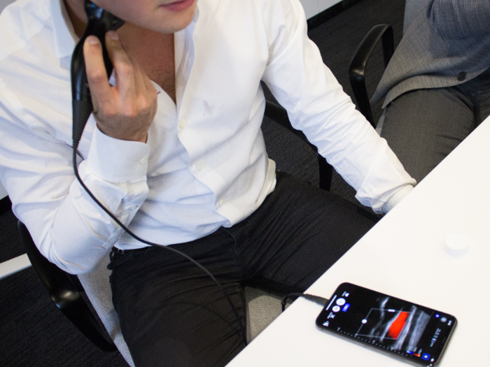
Shahida then showed us the ultrasound on the setting for viewing the carotid artery. Viewing the carotid artery is important, especially since plaque buildup in this area of the body is one of the most common causes of strokes.
The ultrasound can also help guide the insertion of an IV in the central vein, which is often done blindly or with the help of a cardiac visualization technician. "We used to joke that when you were putting a central line in someone, the thing that took the longest was finding the ultrasound machine. Now it's in my pocket," said Martin.
In addition to viewing the ultrasound, the app also lets you annotate and record your scan. You can then save that or send it to a doctor for interpretation.
The data is stored in the cloud after it becomes de-identified. The app is HIPAA compliant, and you can share your data across the globe or across different medical and academic institutions.
Looking forward, the company is working to incorporate parts of the app with electronic medical records, and maybe even bring in artificial intelligence to analyze the ultrasound and tele-diagnose patients for simple maladies.


